


HAWK-超高精度原位蛋白功能定量分析系统
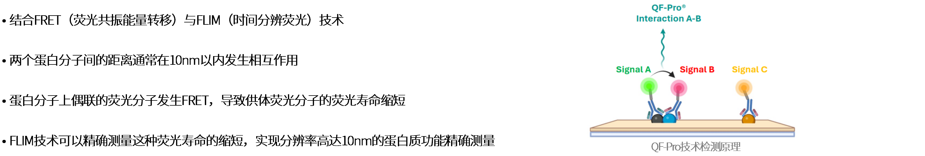
—采用FRET-FLIM技术,在组织切片/固定细胞等样本中,对小于10nm距离的相互作用的蛋白质功能进行精确定量分析
• Hawk Biosystems 公司
HAWK Biosystems位于西班牙,成立于2016年,聚焦空间功能蛋白质组学领域
核心技术 QF-Pro® 平台基于 FRET-FLIM(荧光共振能量转移- 荧光寿命成像显微镜)原理,用于定量分析组织切片和固定细胞等样本中蛋白质功能
2024年推出整体解决方案,具有超高分辨率、高特异性和高灵敏度
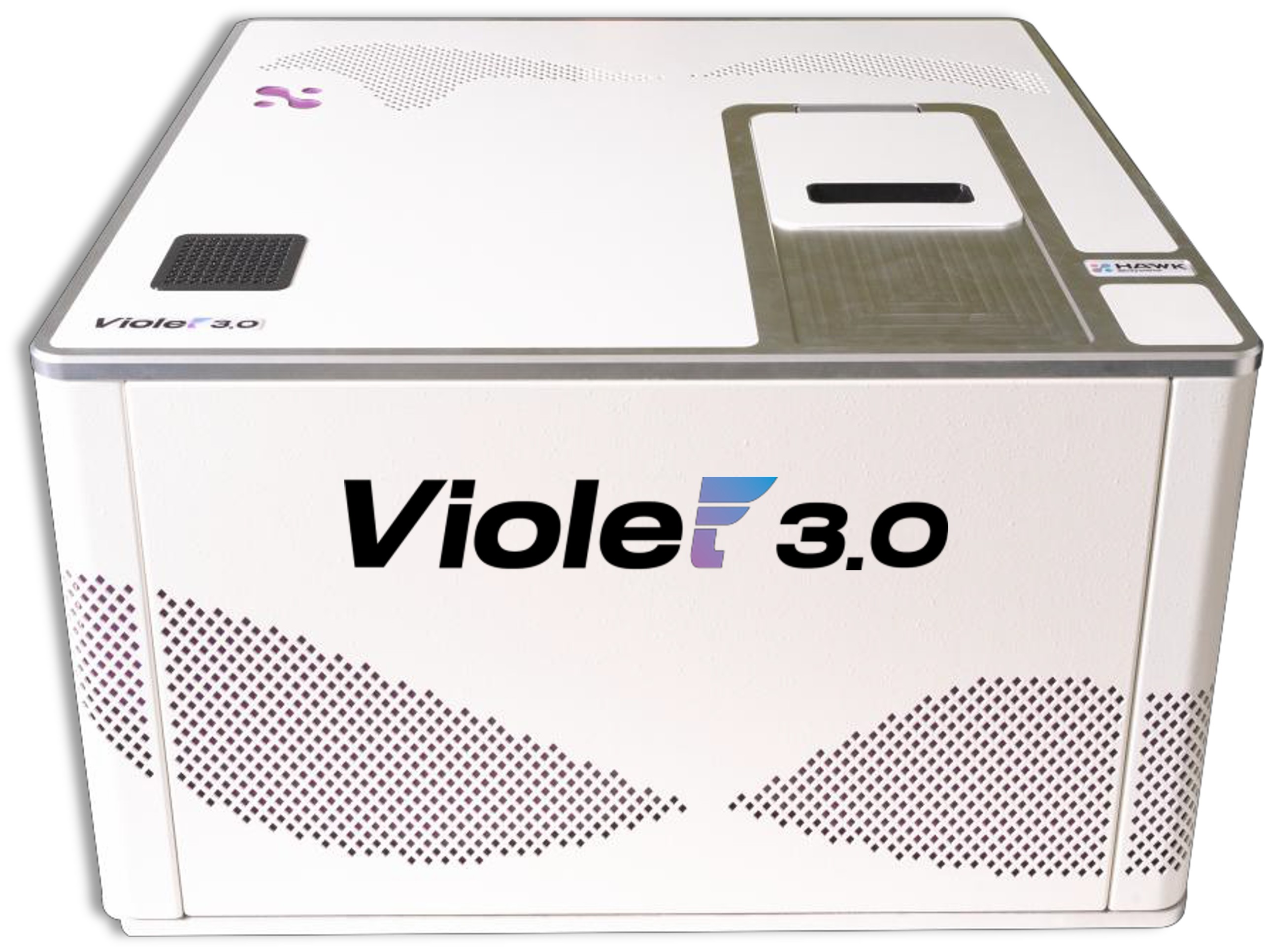
• HAWK-超高精度原位蛋白功能定量分析系统
基于FRET-FLIM检测蛋白质之间相互作用以及蛋白质翻译后修饰等,分辨率可达10nm
特有的信号放大机制有效克服自发荧光,极大提升信噪比
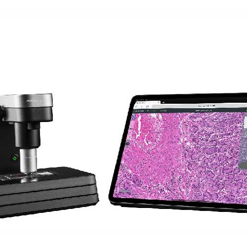
4荧光通道兼容普通免疫荧光染色组织切片/固定细胞成像
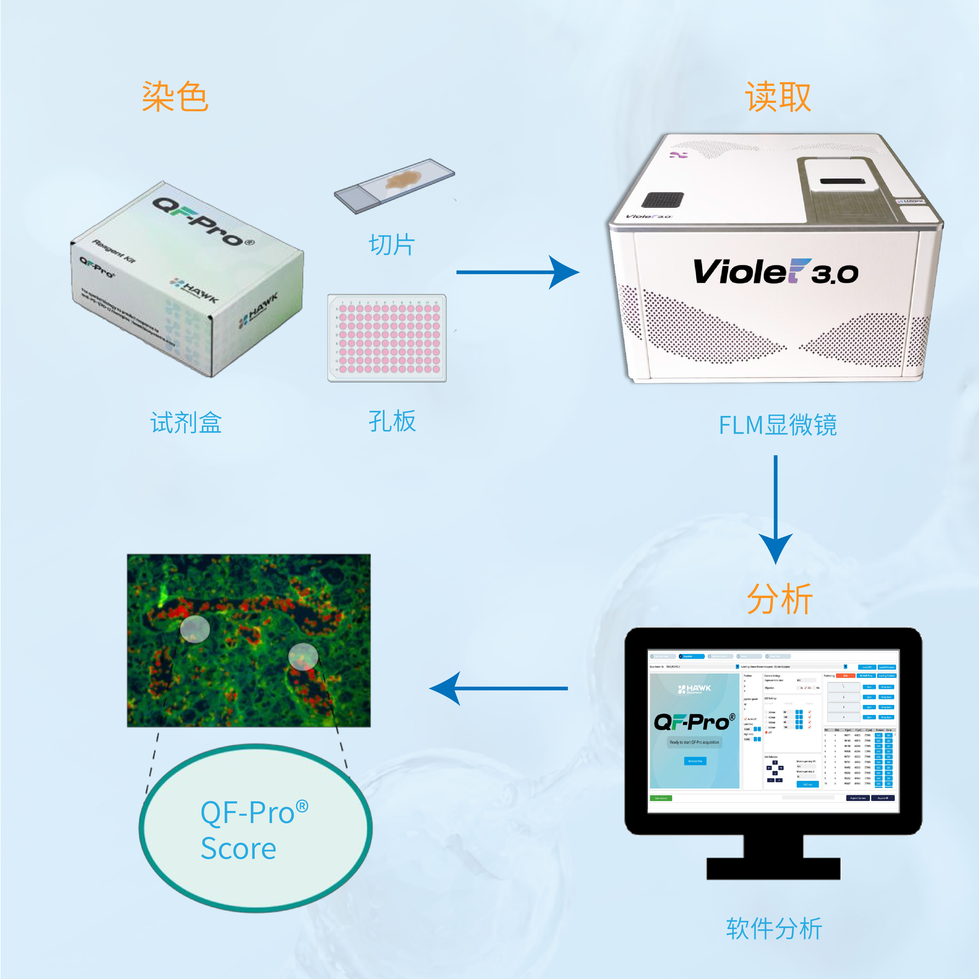
实验流程与普通免疫荧光染色类似,软件使用简单,可以轻松上手

技术原理
产品特点

应用方向

典型案例
案例1 非小细胞肺癌中免疫治疗有效性评估研究背景: PD-L1评分临床诊断中患者是否适合使用PD-1/PD-L1免疫治疗的评判指标,但实践中评判结果并不理想,因此需要找到更有效的评判指标。 研究表明,HAWK的QF-Pro评分是比PD-L1评分更有效的评判指标。
|
|
|
|
|
|
|
案例2 蛋白质与蛋白质相互作用检测研究背景: β-Catenin是一种细胞内蛋白,通过与跨膜蛋白E-Cadherin相互作用发挥功能,HAWK的QF-Pro技术可以识别这两种蛋白是否发生相互作用,并用伪彩显示在免疫荧光染色结果中。
|
|
|
案例3 免疫检查点阻断实验研究背景: TIGIT 与 CD155 的相互作用是肿瘤 “逃避免疫系统攻击” 的关键策略之一。当肿瘤细胞表面的CD155与免疫细胞(如 CD8⁺T 细胞、NK 细胞)表面的TIGIT结合时,会导致免疫细胞杀伤功能失效。因此,用阻断抗体药物阻断CD155与TIGIT结合是一种有效的免疫治疗策略。HAWK可以准确评估阻断抗体阻断CD155与TIGIT结合效果。 |
|
抗体阻断TIGHT和CD155相互作用检测原理
阻断前(左)样本有广泛QF-Pro信号(红色),用抗体阻断后红色信号几乎消失(右)
阻断前后QF-Pro评分对比
|
案例4 蛋白质翻译后修饰研究背景:Akt 蛋白 / 蛋白激酶 B(Akt/PKB)是 “PI3K-Akt 信号通路” 的核心下游分子,其苏氨酸 308 位点(T308)是 Akt/PKB 激活的关键磷酸化位点—— 只有该位点发生磷酸化,Akt/PKB 才能启动激活过程。HAWK的QF-Pro评分可以准确评估不同激活时间下Akt/PKB的激活水平。
|
|
|


发表文献
|
1.Gumuzio et al., 2025. Immune Checkpoint Crosstalk: CTLA-4/CD80 Engagement as a Predictor of Anti-PD-1/PD-L1 Therapy Outcome in NSCLC. Under Review Journal for the Immunotherapy of Cancer (part of the British Medical Journal group). 2.Calleja et al., 2024. Beyond PD-L1: Unraveling the Enigma of Immunotherapy Response in PD-L1 Negative (<1%) NSCLC Patients Through Quantification of PD-1/PD-L1 Engagementin the Tumour Microenvironment. Submitted and Presented as a Poster at the 39th Socienty of Immunotherapy in Cancer (SITC) Congress. 3.Sanchez-Magraner and Gumuzio et al., 2023. Published in the Journal of Clinical Oncology. 4.Miles et al., 2022. Determination of Interactive States of Immune Checkpoint Regulators in Lung Metastases after Radiofrequency Ablation. Published in MDPI Cancers. 5.Sanchez-Magraner et al., 2021. Quantification of PD-1/PD-L1 Interaction between Membranes from PBMCs and Melanoma Samples Using Cell Membrane Microarray and Time-Resolved Förster Resonance Energy Transfer. Published in MDPI Analytica. 6.Sanchez-Magraner and Miles et al., 2020. High PD-1/PD-L1 Checkpoint Interaction Infers Tumor Selection and Therapeutic Sensitivity to Anti-PD-1/PD-L1 Treatment. Published in Cancer Research. 7.Miles et al., 2017. Time resolved amplied FRET identies protein kinase B activation state as a marker for poor prognosis in clear cell renal cell carcinoma. Published in BBA Clinica. 8.Veeriah et al., 2014. High-Throughput Time-Resolved FRET Reveals Akt/PKB Activation as a Poor Prognostic Marker in Breast Cancer. Published in Cancer Research. |
